Introduction
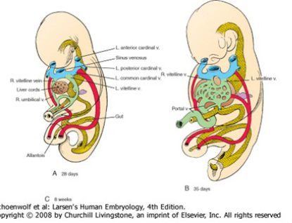
The primitive embryo has 3 venous drainage systems: vitelline system which will drain the GIT and its derivatives, the umbilical system which transport oxygenated blood from the placenta and the cardinal system which drains the body wall, head and neck. All these systems drain into the left and right horns of sinus venous will ultimately drains to the right atrium of the heart.
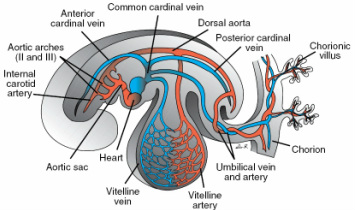
lcoronary section of a late 4th week embryo showing the cardinal, vitelline and umbilical veins
Vitelline System
This forms in the York sac as tinny capillaries which will later join to vitelline plexus of veins and terminate as right and left vitelline veins draining into the right and left horns as the most medial vessels indicated with green in the diagram below. The growing liver bud gradually surrounds the proximal end of these set of vessels with their distal plexuses breaking them down to form the liver sinusoids (blue indications in the 2nd and 3rd diagram in figure below and the liver in orange color).
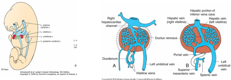
the liver bud invading the superior part of vitelline veins transforming them to sinusoids
Most of the cranial part left vein usually regresses with the left sinus venous (which the ruminate forms the oblique vein of the heart) thus having to develop another vein which will drain the left side of the GIT. The caudal part of the vetilline vein diverse to drain into the now enlarged right vitelline vein by forming a portal system (via a portal vein). The superior part of the right enlarged vitelline vein forms the most proximal part of the inferior veno cava (IVC).
Umbilical system
formation of the portal vein and ductus venous
The umbilical vein in the early embryo is made of 2 veins, the right and left veins which drains into the sinus horn just lateral to the vitelline veins.the liver gradually invades these vessels as it grows sending these vessels to remodeling themselves. In the case of the umbilical vein, the right veins is the one that regresses as the left persist but loses its communication with sinus horns as it forms a new network with the right vitelline vein (indicated by the red vessel in the diagram), which is now superior part of IVC. This connection is a by-pass to IVC containing pure oxygenated blood from the placenta. This by-pass is called the ductus venous.
Cardinal System
The cardinal system as we said earlier drains the whole body wall, head and neck. At an early stage, it consist of an anterior pair and a posterior paired cardinal veins which opens to the heart via right and left common cardinal veins.
Posterior cardinal veins
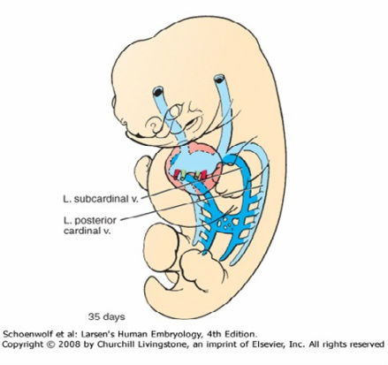
The posterior cardinal veins(Indicated by the lighter blue colour in the diagrams) are supplemented and will be largely replaced by additional pairs: subcardinal and the supracardinal veins.
The subcardinal veins(Seen in the diagrams below deep blue) appear from the base of the posterior cardinals by the end of 6th week, and by the 7th and 8th week, the subcardinal veins become connected with each other via a median anastomosis and to the posterior cardinal vein via a lateral anastomosis. The left subcardinal vein gradually regresses leaving its anastomosis thus the left body wall drained by the right subcardinal vein its anastomotic branches at same time the right subcardinal losses its communication with the posterior cardinal as it develops a new network with segment of the right vitelline vein inferior to the developing liver to form part of the IVC. This part will drain the developing kidney, supra renal gland and the developing gonad initially from its median plexuses.
Posterior cardinal veins
The posterior cardinal veins(Indicated by the lighter blue colour in the diagrams) are supplemented and will be largely replaced by additional pairs: subcardinal and the supracardinal veins.
The subcardinal veins(Seen in the diagrams below deep blue) appear from the base of the posterior cardinals by the end of 6th week, and by the 7th and 8th week, the subcardinal veins become connected with each other via a median anastomosis and to the posterior cardinal vein via a lateral anastomosis. The left subcardinal vein gradually regresses leaving its anastomosis thus the left body wall drained by the right subcardinal vein its anastomotic branches at same time the right subcardinal losses its communication with the posterior cardinal as it develops a new network with segment of the right vitelline vein inferior to the developing liver to form part of the IVC. This part will drain the developing kidney, supra renal gland and the developing gonad initially from its median plexuses.
As the subcardinal veins are undergoing remodeling, the supracardinal veins As seen as purple in the diagrams below)also form also from the base of the posterior cardinal veins to lie in-between the posterior and subcardinal veins giving rise to numerous lateral anastomotic branches to posterior cardinal. As the posterior cardinals’ degenerates, the anastomotic branches of the suprcardinal drains the body wall majorly as intercostal veins having thoracic and abdominal parts which give rise to different components in adult. In the abdominal region, the inferior part of the supracardinal vein degenrates and its remaining segments of the right supracardinal form a segment of IVC just below the kidney. The thoracic part of the supracardinal drains the thoracic wall via serirs of intercostal veins. At the point of communication with the degenerating posterior cardinal vein, the 2 suprcardinal communicate with each other vai a median anastomosis called the accessory hemiazygos vein. The left thoracic supracardinal vein now called the hemiazygos vein soon lose it connection with the reminating left posterior cardinal vein thus with the sinus horns to drain into the right supracardinal vein. The remaining portion of the inferior right supra cardinal vein now called the azygos vein also lose its connection with posterior cardinal vein as it anatomies with segment of the superior veno cava (SVC) which is derived from anterior cardinal vein.
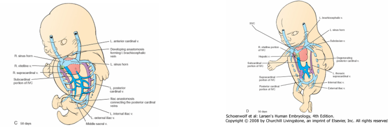
the posterior cardinal vein and supplentary veins giving rise to the IVC
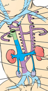
the IVC made up of the vitelline nein in green, subcardinal vein in deep blue, supracradinal vein in purple and the sacral part of IVC with the common, internal and external carotid from posterior cardinal vein in light blue
The right Posterior cardinal vein have lost most of cranial part, only left with a caudal part and its medial anastomosis at that part. Its communication with the caudal part of supracardinal develops into the inferior most (sacral) part of the IVC, the common iliac, it sprout to form the internal and common iliac veins.
Anterior cardinal vein
It is made of right and left cardinal veins which will form veins that drain the upper limb, head and neck.The 2 cardinal veins anastomoses medially to form brachiocephalic vein, as this vein is forming, the left sinus horn is gradually regressing leaving a small part of it that will be the oblique vein of the heart.
The cranial portion of the anterior cardinal becomes the internal jugular as capillary plexus forming in the face will form the external jugular vein.Capillary plexus in the upper limb will form the subclavian vein which opens into the proximal part of the anterior cardinal now right brachiocephalic vein and to the left brachiocephalic vein on the left.
Portion inferior to the junction of thr right and left bracocephalic veins forms the SIV which opens to the right atrum of the heart as the IVC will.
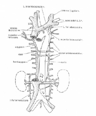
the coollection of the great venous vessels
