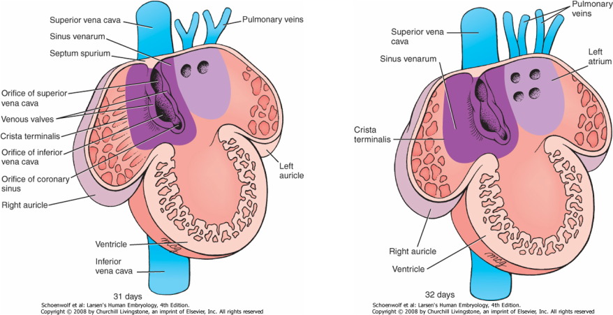
Right atrium
The 2 developing atria have muscular surface: trabecula, before the incorporation of the vessels to each one of it. This muscle is called the pectinate muscle of the atrium.
Sinus venous
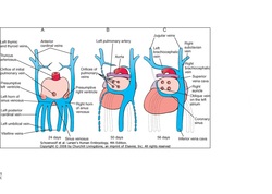
The obliteration of the right umbilical vein and the left vitelline vein
during the fifth week,the sinus horn becomes less important and completely
losing its importance when the left common cardinal obliterate at the 10th week.
Just a small part of it remains which forms the oblique vein of the left atrium
and as the coronary sinus as shown in the diagram.
during the fifth week,the sinus horn becomes less important and completely
losing its importance when the left common cardinal obliterate at the 10th week.
Just a small part of it remains which forms the oblique vein of the left atrium
and as the coronary sinus as shown in the diagram.
The right sinus horn and veins is enlarged greatly as a result of left-to-right shunts of blood thus forming the only communication between the original venous and atrium. As the atrium expands the sinus venous is incorporated into the right atrium to form the smooth-walled part of the right atrium by displacing the trabeculated tissue.The smooth tissue forms part of the atium called the sinus venarum.
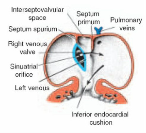
Internally, the sinoatrial oriface is flanked by two valves, the right and left venous valves. Superiorly these two valves meet to form the septum spurium. Note that the left horn opens up underneath the oriface of the right horn
(sinoatrial oriface). Incorporation of the right sinus horns into the atrium will set the fusion of the septum spurium and the developing atrial septum making the superior portion of the right venous valve disappears entirely and the inferior portion to develop into two parts: the valve of the inferior vena cava and the valve of the coronary sinus .
(sinoatrial oriface). Incorporation of the right sinus horns into the atrium will set the fusion of the septum spurium and the developing atrial septum making the superior portion of the right venous valve disappears entirely and the inferior portion to develop into two parts: the valve of the inferior vena cava and the valve of the coronary sinus .
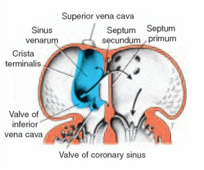
The crista terminalis as shwn in the diagram a ridge of tissue located to the right of the sinoatrial oriface, forms the boundry between the auricle and the sinus venarum dividing line between the original trabeculated part of the right atrium and the smooth-walled part (sinus venarum), which originates
from the right sinus horn.
from the right sinus horn.
The left atrium
The pulmonary veins gradually opens into the left atrium as shown in the diagrams below. the common left and right veins empties and soon gets incorporated into the walls of the left atrium bring the 2 branches of the pulmonary veins on each side to open directly into the left atrium. Just as in the case of the right atrium, the left also loses it trebeculae muscle part to smooth part from the incorporated vessels.The left atrium in an adult life has 4 openings of the pulmonary
veins on its posterior surface .
veins on its posterior surface .