Amazingly the heart starts beating rhythmically at day embryonic day 22 and by day 24 to 25, blood starts flowing thus making it the first functioning organ in humans while much of cardiac development, including remodeling and septation, occurs while the heart is pumping blood. Morphologically, the embryonic heart is first identifiable as a single heart tube composed of contractile myocardium surrounding an inner endocardial (endothelial) tube, with an intervening extracellular matrix.
Endocardial tube
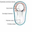
Cardiac progenitor cells are localized within the cranial lateral plate mesoderm (splanchnic layer) on both sides of the embryo extending cranial to the developing brain rostral to the buccopharyngeal membrane forming a cardiac crescent within the precardic cavity . Mesoderm around the cardiac crescent (endocardium) are induced by the underlying pharyngeal endoderm to form cardiac myoblasts.
folding of the embryo
The lateral and craniocaudal folding of the embryo which Commences in the 4th week due to rapid growth of the neural tube leads to fusion of the paired vessels in the lateral mesoderm forming a single endocardial tube while the craniocaudal folding leads to folding of the cardic tube, its surrounding mesoderm and enclosing pericardial space to a position in the thoracic cavity ventral to the invaginated foregut.The folding inverts the orientation of the developing heart relative to the neural structures and the gut.
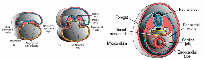
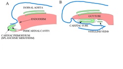
The effect of the lateral folding, A showing the cell clusters,B the paired dorsal aorta formed and the endocardial tubes within its cavity coming to fuse in the midline. the bigger figures shows the fused endocardial tube,surrounding myocardium, pericardial cavity and the mesocardium the 2 dorsal aorta behind the foregut.

transverse section showing the 2 paired vessels forming with relation to other structures http://php.med.unsw.edu.au/embryology/index.php?tittle=file:foster124.jpg
Mesenchymal coverings of the endocardial tube
The fused endocarial tube is now attached to the dorsal side of the pericardial cavity by a fold of mesodermal
tissue: the dorsal mesocardium, this will soon degenerate forming the transverse cardic sinus as it suspended the heart in the pericardic cavity by vessels.
The lateral plate visceral mesoderm proliferates,surrounds the endocardial heart tubes, and develops into the myocardium. During these events, the myocardium thickens and secretes a thick layer of extracellular matrix, rich in
hyaluronic acid, that separates it from the endothelium (endocardium) called the cardiac jelly.
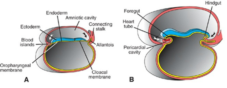
Stages of the crianocaudal folding of the embryo
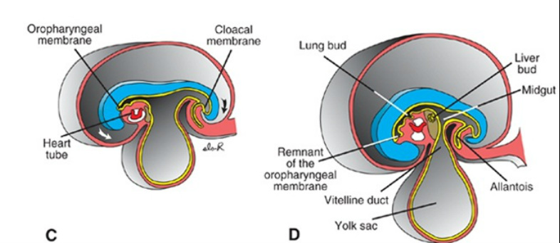
Before and after the cranial folding
this is just an scematic presentation of what happens to the aorta before and after the cranial folding
Looping: bending and rotation
Dilatation and constrictions of the heart, dividing the heart into sections
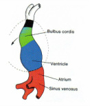
As the endocardial tube folds, it enlongates producing series of constrictions (sulci) which divids the heart tube into sections or expansions: sinus venosus, into which the common cardinal veins, the umbilical veins and the vitelline veins drain; the primitive common atrium; primitive ventricle; and the bulbus cordis, through which blood flows to the paired dorsal aorta and crianlal most is the outflow conus arteriosus/ conus cordis, the truncus arteriosus and the aortic sac.
the endocardial tube now in the thoracic, with all parts formed is thrown into bending and rotationwhich will give its its shape and reposition the great vessels.
the endocardial tube now in the thoracic, with all parts formed is thrown into bending and rotationwhich will give its its shape and reposition the great vessels.
First the endocardial tube becomes C-shaped where the ventral surface forms the right outer Curvature of the C and dorsal surface becoming the inner curvature. It then gradually become S-shape with it distal part moving dorsally to anchor itself on the proximal part of the tube and repositioning the inflow vessels on its posterior surface as the out flow in the ventral aspect.
The part of the atrium anchoring it to the ventricle is will form the right and left auricles of the atria.
The part of the atrium anchoring it to the ventricle is will form the right and left auricles of the atria.
The C and S-shape formation
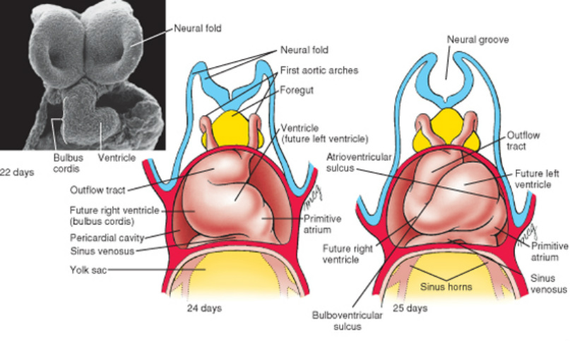
the ventral view of the endocardial looping: at day 24 its C-shaped and an S-shaped at day 25
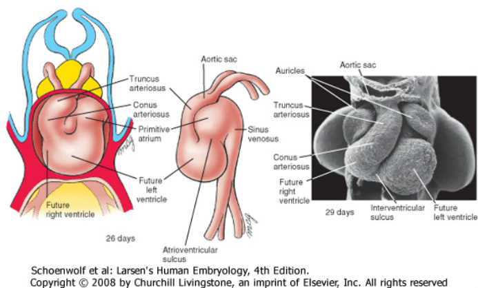
two vental and one lateral view of the looping endocardial tube. already the ventricles and atria are final position. the day 29 shows the anchoring left and right auricles
After looping, the heart tube septates into a four-chambered structure in which the systemic and pulmonarycircuits are distinct.Composing of 2 chambers presently: a single ventricle which is inferior anterior and a single atrium which is superior posterior, therefore it goes into septation (formation of septum) to become 4 chambered structure.
Septation
Enlargement of the heart is mostly with differential growth of myocardium which releases an extracellular matrix in between the myocardium and the endocardium called the cardiac jelly, which directs the growth of the cardiac wall towards the lumen thus initiating the septation. The cardiac jelly alongside the myocardium and migrated neural crest cells contribute in the formation of the cardiac ridges, valves and the skeleton of the heart.
Atrioventicular Septum (Av-septum)
Forms from ridges called the endocardia cushions, originating from midline of anterior, posterior and the lateral walls of the AV-junction to fuse at the midline. This greatly reduces the opening between the two primitive chambers as the communication now in via two openings called the AV canals which are important as it separates the systemic and pulmonary circulations in the adult heart. This opening is initially more to the left side, as the single ventricle is enlarging and dividing into two, the cushion moves towards the right coming to its mid-way position.
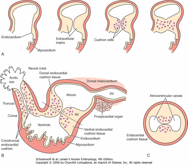
formation of the AV setum or cushion from balloning inwards of ridges formed by myocardium
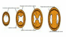
section through the heart illustrating the stages of AV septations as with a result of right and left AV canals
Interatrial Septum
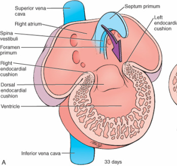
the upper two: scematic illustration of the septum primum at coronary section of the heart
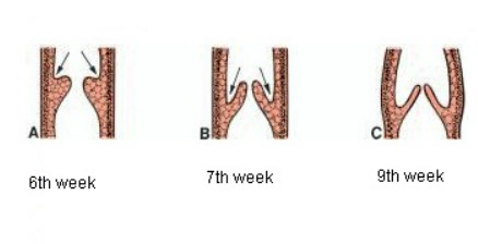
This septum is developing to partition the atrium into right and left atria. Around the 27th day, an ingrowth septum is seen coming from the roof of the atrium almost at the midline as seen in the diapharg represented bt the blue structure. As the atrium expands, the septum gradually grows downward forming a crescent-shaped wedge and narrowing the path as it grows towards the AV cushion, this is called septum or ostimum premium. The narrow path between the septum and the AV cushion is called the premium foreman which allows the flow of blood to the formed left atrium. The septum continuous to grow until it fuses with the cushion and completely closing the foreman but before the foreman is completely obliterated a programmed cell death (apoptosis) occurred at a distal part of the septum closer to the roof creating an opening. This opening is called the foreman secundum as it takes the job of foreman primum.
Another crescent wedge shaped invagination is seen coming also from the roof of the atrium to the right of the septum primum making its way to the Av cushion as shown in the diagrams below, it is indicated by the green while the septum primum is in blue. This is called the septum or ostimum secundum. This grows towards the AV cushion but does not fuse with it, leaving an opening called the foramen oval which allows the flow of blood from right to left atrium.
Books say that this septum is thicker than the septum primum that is reason why septum secundum is rigid while septum primum moves in the fetal heart in opening and shutting the ovals foreman allowing flow of blood from right atrium but preventing reverse action but it is difficult to see in an adult heart as both septa are fused together. You can compare the thickness in thses diagrams.
Books say that this septum is thicker than the septum primum that is reason why septum secundum is rigid while septum primum moves in the fetal heart in opening and shutting the ovals foreman allowing flow of blood from right atrium but preventing reverse action but it is difficult to see in an adult heart as both septa are fused together. You can compare the thickness in thses diagrams.
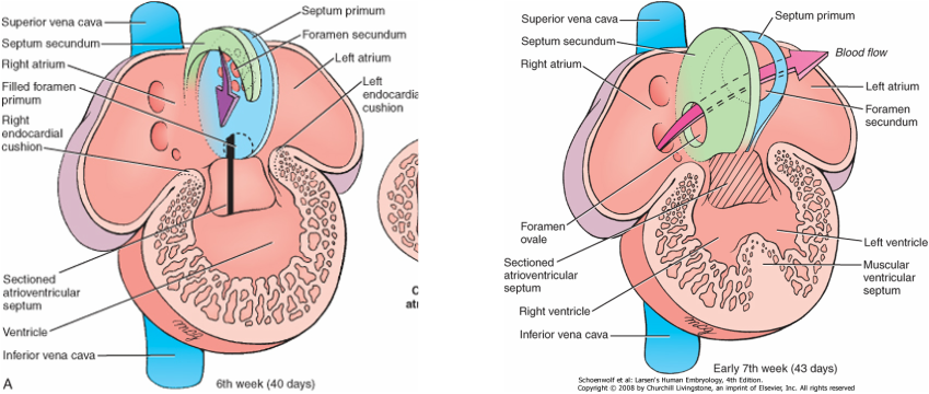
figure A is showing the intiation of the septum secundum as well as the foreman. the2nd figure shows the foreman ovale and the movemnet of setum primum against septum secundum.
Interventicular septum (IV septum)
Formation of this septum takes place between the 4th and 7th week. By mid-4th to 5th week growth and expansion of the embryonic ventricles occurs such that the right ventricle forms by enlargement of the heart tube and incorporation of bulbo cortus to it whereas the left ventricle forms by bulging from the outer curvature of the heart tube thus eventually pulling the AV canals and cushion from the left to the medially as seen in the illustration below.
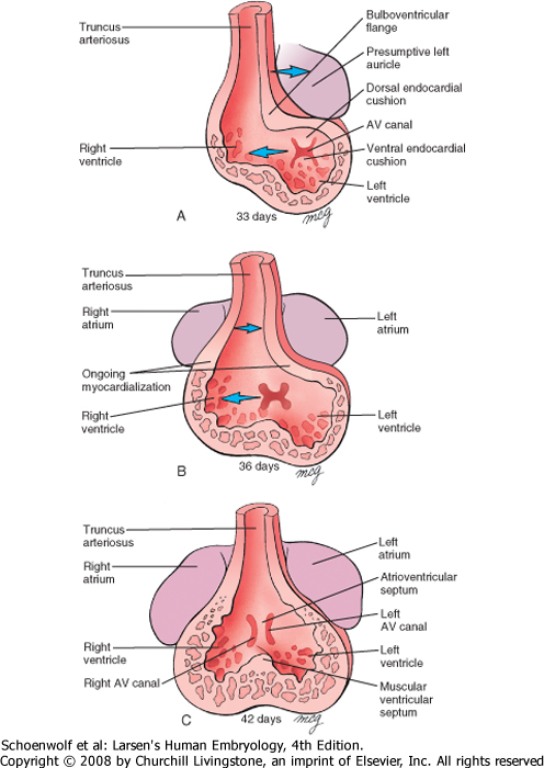
illustration of the partitioning of the ventricles as the AV canals and Av cushion are pulled medially from the left side. an the intiation of the muscular part of IV septum
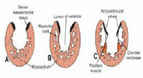
The myocardium thickens forming ridges on the wall of the ventricle called the trabeculae (finger-like projections) which are transformed into fenestrated trabecular and gradually thickens. A ridge is seen on the midline floor of the ventricle growing superiorly towards the AV cushion as both ventricles enlarge forming the IV septum. This septum does not fuse to the AV cushion leaving a blood flow path between the two ventricles called the IV foreman.
The ventral part of IV septum is also thrown into trabeculation in continuation with that of the wall while the dorsal smooth side called the inlet septum which forms a prominent trabeculae called septomarginal trabecular which connects the muscular septum with the forming papillary muscles.
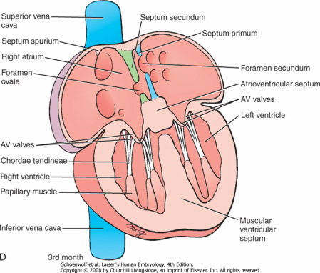
coronary section of the heart, showing the formed foramen oval, Av setum and the incomplete IV septum.the formed Av valve
AV Valves formation
The AV valves are call cusp which are firmly attached to the rim of both AV canals. This forms between 5th and 8th week from adjacent endocardia cushions tissues as it sends down a muscular connection (via the ventricular trabeculae) to the walls of the ventricles. Thin strands of cells-muscular cords remain from the degeneration of the inferior muscular part called the chordea tendineae is holding the cusp as small hillocks of myocardium called papillary muscle roots it to the walls of the ventricles. The left ventricle has 2 valves: mitral or bicuspid (anterior and posterior cusp) while the right has 3: tricuspid valve (same as left plus an IV septum cusp).
Septation of the outflow
The outflow composes of the distal part of bulbus cordis and Truncus arteriosus. The cTruncus arteriosus l portion of the heart tube, initially on the right side of the pericardial cavity, shifts gradually to a more medial position. Two ridges opposing each other form on the wall of the outflow tract which gradually fuses in the midline partitioning the track and this septum is called the conotruncal or aorticopulmonary septum. Unlike in the case of the other septa, this is not moving straight rather in a spiral form along the walls of this tract so that the right ventricle connects with the feature pulmonary trunk( the blue are in the diagram below) which is located behind the aorta (the red arrow movenment as in diagram below) and the left ventricle connected to the aorta. Myocardium flows into this septum providing musculature as the trucks divides into two vessels.
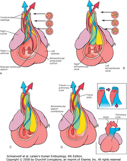
As aorticopulmonary septum is forming, the muscular part of the IV septum we have discussed earlier continou it journey to attach to the ventricular surface of the IV cushion and the inferior part of aorticopulmonary septum which is membranous thus
obliterating the IV foreman and as well completely separating the systemic and pulmonary out flows as seen in the 2 diagrams of this figure.
obliterating the IV foreman and as well completely separating the systemic and pulmonary out flows as seen in the 2 diagrams of this figure.
Semilunar valves
These are valves of the out flow tracks, preventing inflow from the out flow tract to the ventricles. They develop same time as aorticopulmonary septum developing as small tubercles found on the main truncus swellings.One of each pair is assigned to the pulmonary and aortic channels, respectively and third tubercle appears in both channels opposite the fused truncus swellings.Gradually the tubercles hollow out at their upper surface, forming the semilunar valves.
You can watch an amination of tdevelopment of the heart on http://www.youtube.com/watch?v=OArR67aFze0
