Development of the aortic arches
Introduction
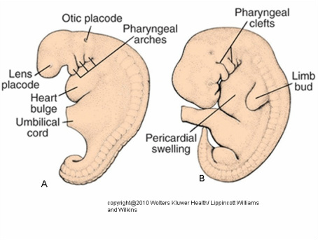
The late 4th week, some elevations are visible on the both side of the neck region of the embryo, this are called the pharyngeal or bronchial pouches.
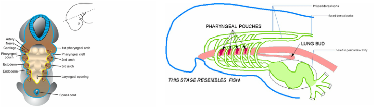
coronary section throught the pharyngeal apparatus showing layers. the structures forming from the mesodermal layer
A coronary section through this area will show 5 paired arches each compose of an ectoderm, mesoderm and endoderm. We are not going to talk about the whole bronchial apparatus but just to let us have a peek of where our aortic arches are coming from. Each bronchial arch mesoderm gave rise to the aortic arch thus our five paired aortic arches.The 5 paired arches connect with the aortic sac of truncus arterosis of the heart that we have discussed earlier.
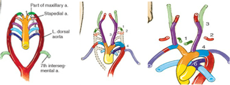
In humans, the 5 paired are: 1,2,3,4 and 6 arches are seen actually as they are seen as in the fihure above . The 5th doses not develop and if it does it degenerates almost immediately. The 5 paired arches are not seen all at one time in humans as most of our figures illustrate, the first and second arches almost completely transform before the others are formed.
The first arch is seen about the time of the cranial folding as it attaches to the right and left dorsal aorta. The left and right dorsal aorta fuses at the end of the 4th week from the 4th thoracic to the 4th lumbar region thus becoming just a single vessel.
The first arch is seen about the time of the cranial folding as it attaches to the right and left dorsal aorta. The left and right dorsal aorta fuses at the end of the 4th week from the 4th thoracic to the 4th lumbar region thus becoming just a single vessel.
As the first arch regresses leaving a very small ruminate which forms the maxillary artery the second arch is being formed. About the 28th day, the 3rd and 4th arches appears as the 2nd regresses leaving a small part which will form the stapedial artery (artery to stapes bone of the ear).
The 3rd arch will give rise to: left and right proximal end of the internal carotid artery and left and right the common carotid artery. The distal end of the internal carotid artery is from aortic sac while the external carotid is from the dorsal aorta both parts superior to point of 3rd arch connection.
By the 7th week, the short right infused dorsal aorta will lose it connections with the single dorsal aorta as well with the 6th arch but remain connected to the 4th and the 7th intersegmental artery to form the right subclavian artery. The region of the aortic sac where the 4th arch attaches forms the right brachiocephalic artery. The left 4th arch will remains connected with the aortic sac and left short infused segment of dorsal aorta to become the arch of aorta as the left 7th intersegmental artery give rise to the left subclavian artery.
By the 7th week, the short right infused dorsal aorta will lose it connections with the single dorsal aorta as well with the 6th arch but remain connected to the 4th and the 7th intersegmental artery to form the right subclavian artery. The region of the aortic sac where the 4th arch attaches forms the right brachiocephalic artery. The left 4th arch will remains connected with the aortic sac and left short infused segment of dorsal aorta to become the arch of aorta as the left 7th intersegmental artery give rise to the left subclavian artery.
the transformation of the aortic arches to arch of aorta and the great arterial vessels
Also at the 7th week, 2 short vessels are seen connecting to the 6th : pulmonary arteries, one to each arch as the right arch losses it connection with the right dorsal. While the left arch maintains it attachment with the dorsal aorta at same time with its new connectio . Connection of the 6th arch with the dorsal aorta on the left side is called the ductus arteriosus.
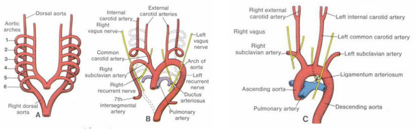
The figure above shows the transformation of the aortic arches with relation to right and left the recurrent laryngeal of vagus nerve. B above is of a fetus while C is of an adult. The persistance of the the connection of the left 6th arch ( forming the ductus arterosus) stationed the left recurrent laryngeal nerve between the arch of aorta and the ligament arteriosus in a adult.
